

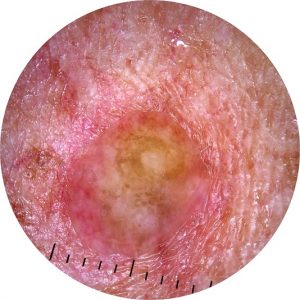
What would you do? A 64-year-old male presents with a cyst on his back & has a personal history of basal cell carcinoma & a family history of melanoma.
-1.jpg)
HealthCert Education
In this case discussion from Dr Heather Lawson, we look at a 64-year-old male patient with a personal history of basal cell carcinoma 10 years ago and a family history of a father with melanoma.
The patient presented to Dr Lawson’s clinic for the first time after being referred by his family physician for removal of a cyst on his back. The cyst had been growing over the past six months.
When Dr Lawson examined the lesion, she felt it was more likely to be solid than cystic, so she excised it in its entirety, different from how she excises cysts (whereby she cuts a small ellipse in the center of the cyst). The lesion was solid.
What are your thoughts on the potential diagnosis?

Update
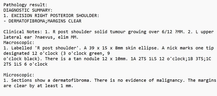
Here is the pathology report:
A note from Dr Heather Lawson:
I really thought this was a melanoma. I have seen a 2cm thick spindle cell melanoma that looked like a cyst to two of the patient’s GPs, resulting in a 5 month delay in excision. I have researched that Dermatofibromas can be up to 15mm wide, but I have never seen one like this before. I did ask the pathologist to recheck, and the diagnosis was the same.
-1.jpg)
-1.jpg)
-1.jpg)